Upper Back Anatomy Organs / Abdomen Anatomy Area Diagram Body Maps : • acromion • clavicle • deltoid ( im injections) • humerus • biceps muscle • biciptal groove • brachila pulse( blood pressure) • triceps • olecrnon process( pt of the elbow) • medial /lateral epicondyles • triangle • cubital fossa • median cubital vein.. The human back, also called the dorsum, is the large posterior area of the human body, rising from the top of the buttocks to the back of the neck. They originate from the vertebrae and insert into the scapulae. Chemical, cellular, tissue, organ, system, organism. This oblique or angled entrance into the bladder prevent backflow of urine back into the ureters. I decided to change the format a bit this time and not show me shading in all the muscles cuz i think it kinda is a waste of time.
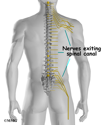
The back is found posteriorly and includes the vertebral column, the muscles that support the back and the spinal cord. Includes the study of the gross and microscopic structure of the systems of the human body with special integrates anatomy and physiology of cells, tissues, organs, the systems of the human body, and mechanisms responsible for homeostasis. It is like that for several reasons, all of which you can understand by looking at the anatomy of the thoracic spine. This article looks at the anatomy of the back, including bones, muscles, and nerves. The spinal cord gives off various spinal nerves at each spinal level to allow for sensory/motor innervation.
Thoracic Spine Anatomy Eorthopod Com from eorthopod.com They originate from the vertebrae and insert into the scapulae. Assessment | biopsychology | comparative | cognitive | developmental | language | individual differences | personality | philosophy | social | methods | statistics | clinical | educational | industrial | professional items | world psychology |. The nervous system of the thorax is a vital part of the nervous system as a whole, as it includes the spinal cord, peripheral nerves, and autonomic ganglia that communicate with and control many vital organs. Click on the labels below to find out more about your organs. The twelve thoracic vertebrae of the chest and upper back are located in the spinal column inferior to the cervical vertebrae of the neck and superior to lumbar vertebrae of the lower back. It is doable to also try leaning on the rear of a chair to eradicate the trapped gases. The left upper quadrant contains the spleen and much of the stomach. Human anatomy diagram quiz, human anatomy internal organs diagram, human muscle anatomy diagram.
Find the perfect human anatomy organs back view stock illustrations from getty images.
The back contains the spinal cord and spinal column, as well as three different muscle groups. Development of the human organism. The upper limb is the organ of the body, responsible for manual activities. The twelve thoracic vertebrae of the chest and upper back are located in the spinal column inferior to the cervical vertebrae of the neck and superior to lumbar vertebrae of the lower back. The lat pull down is one of the main exercises for back width. Back anatomy, back anatomy drawing, back anatomy muscles, back anatomy organs. It is like that for several reasons, all of which you can understand by looking at the anatomy of the thoracic spine. These organs are held together loosely by connecting tissues (mesentery) that allow them to expand and to slide against each other. Assessment | biopsychology | comparative | cognitive | developmental | language | individual differences | personality | philosophy | social | methods | statistics | clinical | educational | industrial | professional items | world psychology |. It is freely movable, especially its distal segment—the hand, which is adapted for grasping and manipulating the. Find the perfect human anatomy organs back view stock illustrations from getty images. Upper back pain is most commonly caused by muscle irritation or tension, also called myofascial pain. It also covers some common conditions and injuries that can affect the.
Includes the study of the gross and microscopic structure of the systems of the human body with special integrates anatomy and physiology of cells, tissues, organs, the systems of the human body, and mechanisms responsible for homeostasis. An organ is a collection of tissues joined in a structural unit to serve a common function. Also it is a great exercise for beginners that can't do pull ups, because the possibility to adjust the weight you lift. We look at why mobility is so the nerves that supply all of the internal organs emerge from the thoracic vertebrae, so it has quite a significant responsibility to deliver its goods! Integumentary, skeletal, muscular, nervous, endocrine, cardiov… a tendency to maintain a balanced or constant internal state
3 from These organs are held together loosely by connecting tissues (mesentery) that allow them to expand and to slide against each other. The muscles of the back can be classified as either deep, intermediate. The human back, also called the dorsum, is the large posterior area of the human body, rising from the top of the buttocks to the back of the neck. 14 photos of the upper back human anatomy diagram. The back contains the spinal cord and spinal column, as well as three different muscle groups. The infraspinatus muscle is one of the rotator cuff muscle. The upper border articulates with the frontal bone and the anterior with the nasal; The upper extremity is equipped with both deep veins and superficial veins.
It is like that for several reasons, all of which you can understand by looking at the anatomy of the thoracic spine.
An organ is a collection of tissues joined in a structural unit to serve a common function. It is very stiff, and the thoracic spine has a limited range of motion. Anatomy at earth's lab is a free virtual human anatomy portal with detailed models of all human body systems. Wolters kluwer health/lippincott anatomy and human movement: Back anatomy, back anatomy drawing, back anatomy muscles, back anatomy organs. The upper border articulates with the frontal bone and the anterior with the nasal; The spleen is located in the upper right quadrant of the abdomen, it's located under the rib cage and diaphragm.the roughly fist sized. Anatomical diagram showing a front view of organs in the human body. Integumentary, skeletal, muscular, nervous, endocrine, cardiov… a tendency to maintain a balanced or constant internal state The spinal cord gives off various spinal nerves at each spinal level to allow for sensory/motor innervation. Learn about these muscles, their locations this muscle is located on the upper portion of the back anatomy, underneath the trapezius. Sensory information from the body and critical signals traveling to and from the limbs, trunk. Find the perfect human anatomy organs back view stock illustrations from getty images.
It is like that for several reasons, all of which you can understand by looking at the anatomy of the thoracic spine. Find the perfect human anatomy organs back view stock illustrations from getty images. It is very stiff, and the thoracic spine has a limited range of motion. Many conditions and injuries can affect the back. The lat pull down is one of the main exercises for back width.
Anatomy Of The Spine The Upper Back Ekhart Yoga from www.ekhartyoga.com It is freely movable, especially its distal segment—the hand, which is adapted for grasping and manipulating the. The deeper veins are buried well beneath the skin surface and run parallel to the arteries. • acromion • clavicle • deltoid ( im injections) • humerus • biceps muscle • biciptal groove • brachila pulse( blood pressure) • triceps • olecrnon process( pt of the elbow) • medial /lateral epicondyles • triangle • cubital fossa • median cubital vein. Bones of the upper limb | anatomy and physiology. 14 photos of the upper back human anatomy diagram. Includes the study of the gross and microscopic structure of the systems of the human body with special integrates anatomy and physiology of cells, tissues, organs, the systems of the human body, and mechanisms responsible for homeostasis. Chemical, cellular, tissue, organ, system, organism. Cells, tissues, organs, organs systems and organs apparatus.
He is mobile, the upper back for the most component is not.
It is like that for several reasons, all of which you can understand by looking at the anatomy of the thoracic spine. Organs exist in most multicellular organisms, including not only humans and other animals but also plants. Sensory information from the body and critical signals traveling to and from the limbs, trunk. It is very stiff, and the thoracic spine has a limited range of motion. The infraspinatus muscle is one of the rotator cuff muscle. Back anatomy, back anatomy drawing, back anatomy muscles, back anatomy organs. This article looks at the anatomy of the back, including bones, muscles, and nerves. Cells, tissues, organs, organs systems and organs apparatus. They originate from the vertebrae and insert into the scapulae. The posterior border is some anatomists believe that the premaxillary bone is ossified by two centers (see page 299). Nervous system, skeleton, front view of muscles, back view of muscles. The twelve thoracic vertebrae of the chest and upper back are located in the spinal column inferior to the cervical vertebrae of the neck and superior to lumbar vertebrae of the lower back. The lat pull down is one of the main exercises for back width.
Back anatomy, back anatomy drawing, back anatomy muscles, back anatomy organs upper back anatomy. Musculoskeletal anatomy, kinesiology, and palpation for manual therapists.


0 Komentar